
 EN
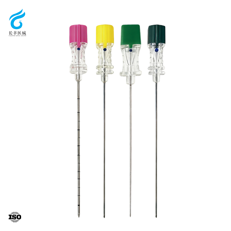
Root Causes of Spinal Anesthesia Failure Linked to Spinal Anesthesia Needle Performance
Operator technique errors: Loss of tactile feedback, incorrect angle, or depth misjudgment
Successful spinal anesthesia hinges on precise needle placement—yet operator technique errors remain a leading cause of failure. Palpation-based identification of the correct intervertebral level is unreliable in up to 64% of cases, particularly in patients with elevated BMI or spinal deformities. This increases the risk of insertion at an unintended interspace, where the dural sac may be absent or the approach angle suboptimal. Thinner needles—often selected to reduce post-dural puncture headache (PDPH)—frequently fail to transmit the characteristic dural “click,” depriving the provider of critical tactile confirmation. Without this feedback, the needle may be advanced too far (risking nerve trauma or intravascular injection) or withdrawn prematurely (failing to achieve CSF return). Depth misjudgment is further compounded in patients with obscured bony landmarks, where first-attempt success rates decline significantly. While real-time imaging guidance can mitigate these issues by enabling precise angle control and depth verification, overreliance on tactile cues alone introduces variable and preventable risk.
Device-related limitations: Needle bevel geometry, stiffness, and tip design affecting CSF flow and dural penetration
The physical properties of the spinal anesthesia needle directly influence procedural reliability. Needle stiffness governs trajectory fidelity: thinner, more flexible needles are prone to deflection off the midline path—prolonging attempts or yielding false-negative loss-of-resistance findings. Bevel geometry determines how tissue is engaged; sharp cutting bevels may core a skin or ligament fragment that occludes the lumen, delaying or preventing CSF aspiration. In contrast, pencil-point designs produce slower CSF flow—a feature associated with lower PDPH but sometimes misinterpreted as failed placement. Tip design also modulates the dural puncture sensation: atraumatic tips yield a less distinct pop, increasing uncertainty about intrathecal entry. When column strength is insufficient, advancing force dissipates into soft-tissue bending rather than controlled dural penetration—especially problematic in patients with calcified ligaments. These device-related variables don’t merely affect comfort or preference; they directly impact block adequacy and the need for conversion to general anesthesia.
Ensuring Spinal Anesthesia Needle Patency: Occlusion Mechanisms and Verification Protocols
Patency is a fundamental prerequisite for reliable spinal anesthesia. A blocked needle before or during injection risks incomplete blockade, wasted drug, and avoidable patient distress. Systematic checks targeting the most common occlusion mechanisms—and confirming free flow—are non-negotiable components of safe practice.
Common occlusion sources — tissue plug, blood reflux, and CSF viscosity — and their prevention
Three mechanisms account for the majority of flow obstructions. A tissue plug forms when a cutting-tip needle cores a fragment of skin or ligament during insertion—occurring in up to 15% of punctures (2023). Using an introducer needle reduces tissue coring by approximately 70% (2020). Blood reflux, often from traversing the epidural venous plexus, can clot inside the lumen; gentle aspiration before injection—and flushing with preservative-free saline if blood is noted—prevents sustained occlusion. Elevated CSF viscosity, though rare, may impede flow in very small-gauge needles; selecting a 25G or larger needle for patients with suspected hyperviscosity mitigates this risk, while avoiding contact between the needle tip and non-sterile surfaces preserves lumen integrity.
Stepwise patency checks: Free-flow CSF aspiration, low-resistance saline test, and real-time pressure monitoring
Begin by confirming free-flowing CSF aspiration after entering the subarachnoid space: withdraw the stylet and observe for a clear, steady drip. Absence of flow suggests a tissue plug—rotating the needle 90° often restores it. Next, attach a syringe filled with preservative-free saline and apply gentle pressure; minimal resistance confirms patency, while high resistance signals obstruction. Finally, connect a pressure transducer to the needle hub. Real-time pressure monitoring displays a pulsatile waveform synchronized with arterial pulsations; a flat or dampened trace indicates blockage and mandates repositioning or needle replacement before drug administration (2024). Performed sequentially in under 60 seconds, this protocol dramatically reduces unplanned repeat punctures and supports first-pass success.
Optimizing Spinal Anesthesia Needle Selection: Atraumatic vs. Conventional Designs by Clinical Scenario
Evidence-based comparison: Pencil-point (e.g., Whitacre, Sprotte) vs. Quincke needles in block success and complication profiles
Clinical evidence strongly supports pencil-point needles for reducing post-dural puncture headache (PDPH): a 2023 meta-analysis found rates of 1–3% with non-cutting tips versus 8–12% with cutting-tip designs in young, healthy patients. This benefit stems from the atraumatic bevel’s ability to separate—not transect—dural fibers. Block success rates, however, are comparable when both types are used by experienced clinicians. That said, the subtler dural feedback and slower CSF flashback of pencil-point needles correlate with higher technical failure among trainees and increased redirection attempts. In contrast, cutting-tip needles deliver superior tactile feedback and faster CSF return—advantages that shorten procedural time and improve first-pass accuracy, especially in time-sensitive or high-acuity settings.
Context-specific guidance: When conventional spinal anesthesia needle designs remain preferred — obstetrics, high BMI, or difficult anatomy
Conventional cutting-tip needles retain clear clinical utility in specific scenarios. In obstetrics—particularly emergency cesarean delivery—the rapid, unambiguous confirmation of CSF return can be life-saving. In patients with high BMI, increased tissue depth attenuates the subtle resistance change of pencil-point entry; the distinct “pop” of a cutting bevel helps avoid dural tenting and inadvertent over-insertion. For challenging anatomies—including scoliosis or prior spinal surgery—the ability to redirect a cutting needle without enlarging the dural hole preserves dural integrity, reduces CSF leakage, and maintains directional control for successful block placement.
Serious Complications Directly Tied to Spinal Anesthesia Needle Use
Dural puncture severity and post-dural puncture headache risk modulated by needle size, bevel orientation, and insertion technique
Dural injury severity—and subsequent PDPH risk—is directly shaped by needle characteristics and technique. Accidental dural puncture leads to PDPH in over 50% of cases (2022). Smaller-gauge needles (e.g., 27G) consistently lower risk by minimizing the dural defect. When using a cutting needle, orienting the bevel parallel to the longitudinal axis of the spine promotes separation—not transection—of dural fibers, further reducing leakage. Multiple insertion attempts or improper angulation compound trauma; thus, meticulous single-pass technique remains foundational to minimizing PDPH, especially in high-risk groups like parturients.
Rare but critical events: Needle breakage, epidermoid tumor implantation, and prevention through device integrity checks and proper handling
Though rare, needle breakage and epidermoid tumor implantation represent serious, preventable complications rooted in device integrity and handling. Before each use, visually inspect the spinal needle for cracks, tip burrs, or signs of fatigue—and discard any compromised device. Breakage most commonly occurs when excessive force is applied against bony resistance; advancement must always be steady and controlled. Epidermoid tumors arise from epidermal tissue carried into the subarachnoid space—eliminated entirely by using an introducer needle and avoiding tissue coring. Manufacturer-recommended handling, routine visual inspection, and gentle, deliberate insertion constitute the essential, evidence-informed prevention strategy.
Frequently Asked Questions (FAQ)
Why might spinal anesthesia fail?
Spinal anesthesia can fail due to operator technique errors, device limitations like needle design affecting CSF flow, or issues like needle occlusion caused by blood reflux, tissue plugs, or high CSF viscosity.
How can I ensure spinal anesthesia needle patency?
Systematic checks include free-flowing CSF aspiration, testing resistance with preservative-free saline, and real-time pressure monitoring to confirm needle patency before drug administration.
What needle type reduces post-dural puncture headaches?
Pencil-point needles are most effective at reducing post-dural puncture headache risk, with studies showing rates between 1–3% compared to 8–12% with cutting-tip needles.
Are cutting-tip needles better for any specific scenario?
Yes, cutting-tip needles are preferred in scenarios like obstetrics, high BMI, or patients with difficult anatomies due to their faster CSF return and clearer tactile feedback.
How can I minimize serious complications with spinal anesthesia needles?
Prevent complications like needle breakage or epidermoid tumor implantation by inspecting the device for integrity, handling it gently, and using an introducer needle to avoid tissue coring.
Table of Contents
- Root Causes of Spinal Anesthesia Failure Linked to Spinal Anesthesia Needle Performance
- Ensuring Spinal Anesthesia Needle Patency: Occlusion Mechanisms and Verification Protocols
- Optimizing Spinal Anesthesia Needle Selection: Atraumatic vs. Conventional Designs by Clinical Scenario
- Serious Complications Directly Tied to Spinal Anesthesia Needle Use
- Frequently Asked Questions (FAQ)