
 EN
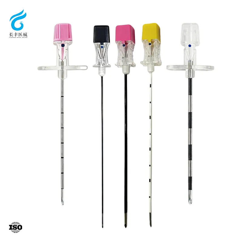
Anatomic Foundations: How Pediatric Spine Development Guides Needle Selection
Successful regional block placement in children relies on a thorough understanding of the dynamic pediatric spine. Unlike the mature adult column, the infant and child spinal axis undergoes significant transformation that directly influences where and how a pediatric anesthesia needle is safely advanced.
Vertebral Level Variability and Dural Sac Termination Across Age Groups
The conus medullaris and dural sac do not remain fixed; their positions relative to the vertebral bodies shift cephalad throughout early childhood. At birth, the dural sac terminates as low as the S3–S4 interspace, while the conus usually ends around L3 (Dalens 2014). Over the first few years, differential growth of the vertebral column and spinal cord causes an apparent ascent: by age 5, the dural sac’s end typically reaches S1–S2, and the conus stabilizes at L1–L2, mirroring adult proportions. This variability creates a narrower margin of safety in infants: a lumbar puncture or epidural placement at a seemingly “safe” L4–L5 interspace can still risk dural puncture or cord injury in the very young. Relying on the iliac crest line (Tuffier’s line), which often crosses L4–L5 in children, helps—but is less reliable before walking age. Practitioners must therefore select the most caudal safe interspace—commonly L4–L5 or L5–S1 for spinal anesthesia—and always angle the pediatric anesthesia needle slightly cephalad to respect the still-low termination. Routine pre-procedural ultrasound scanning can confirm the conus position and substantially reduce needle misplacement in these age groups (Sury et al. 2016).
Skin-to-Dura Distance and Its Impact on Pediatric Anesthesia Needle Length
Age-specific tissue dimensions determine the minimum needle length required to reach the subarachnoid or epidural space without overshooting. The distance from the skin surface to the dura mater follows a nonlinear increase: in term neonates, the median skin-to-dura depth is only about 1.3–1.6 cm (Polaner 2017). By toddlerhood (1–3 years), this distance grows to roughly 2.0–2.5 cm, and by adolescence it approaches 3.0–4.0 cm, comparable to adults. These shallow depths mean that standard 90 mm spinal needles designed for adults introduce unnecessary risk of excessive insertion, potentially leading to ventral epidural hematoma or unintended intrathecal catheter placement. Weight- or body surface area–based formulas can predict needed depth within ±5 mm, but ultrasound measurement provides greater accuracy. Most pediatric anesthesia needle protocols therefore recommend that for infants under 5 kg, a 25–30 mm styletted needle is adequate, while a 50 mm needle is sufficient for most children under 30 kg. Selecting a shorter needle based on measured skin-to-dura distance, rather than a one-size-fits-all approach, reduces the volume of tissue trauma and the likelihood of accidental dural puncture with a longer, unwieldy tool.
Needle Design Matters: Cutting vs. Pencil-Point for Pediatric Anesthesia Needle Safety
PDPH Risk Reduction with 27G Pencil-Point Needles in Children and Adolescents
Post-dural puncture headache (PDPH) arises from persistent cerebrospinal fluid leakage after the dura is breached during lumbar puncture. The needle tip design is the principal controllable factor determining the magnitude of this leak. A cutting needle, such as a Quincke type, transects dural fibers cleanly, leaving a crescentic defect that seals slowly. In contrast, a pencil-point needle spreads the fibers apart, creating a small slit that closes promptly once the needle is withdrawn. A Cochrane systematic review confirmed that pencil-point needles significantly reduce the odds of PDPH compared with cutting needles (Arevalo‑Rodriguez 2017). In pediatric studies, the 27‑gauge (G) pencil-point needle consistently yields a PDPH incidence below 1%, making it the preferred choice for spinal anesthesia in children. Adolescents, whose larger dural sac and predominantly upright posture elevate baseline risk, gain the most from this design. Although the 27G pencil‑point needle requires an introducer for skin puncture, the added step does not compromise safety. The combination of ultra‑fine gauge and a non‑cutting tip is a reliable, evidence‑backed strategy to virtually eliminate PDPH in young patients.
Current Evidence-Based Guidelines for Pediatric Anesthesia Needle Use
Contemporary guidelines from the American Society of Regional Anesthesia and Pain Medicine (ASRA) and the European Society of Regional Anaesthesia (ESRA) mandate the preferential use of pencil‑point spinal needles over cutting designs in children. The ASRA 2021 practice advisory specifies that a non‑cutting, pencil‑point tip and the smallest gauge consistent with procedural needs—typically 25G or 27G for patients under 30 kg—should be employed. An introducer is strongly recommended to prevent tissue coring and allow precise control of the fine‑gauge pencil‑point needle. Surveys of pediatric anesthesiologists reflect this shift: while 22G Quincke needles were once common, over 80 % of practitioners now select pencil‑point needles for spinal blocks. Ultrasonographic measurement of skin‑to‑dura distance further guides length choice, minimizing inadvertent bone contact and failed attempts. These authoritative recommendations make clear that tip design is a critical safety measure, not a minor preference. Adhering to guidelines—non‑cutting tip, small gauge, and ultrasound‑assisted length selection—ensures that every lumbar puncture is as atraumatic as possible, reducing both immediate and delayed complications.
Optimizing Pediatric Anesthesia Needle Specifications: Gauge, Length, and Entry Site
Age- and Weight-Based Recommendations for Pediatric Anesthesia Needle Gauge and Length
Selecting the correct pediatric anesthesia needle gauge and length depends on the child’s age and weight to avoid neural injury while ensuring reliable dural puncture.
| Age Group | Weight (kg) | Gauge | Needle Length (mm) | Entry Site |
|---|---|---|---|---|
| Preterm neonate | <2.5 | 27G pencil‑point | 25–30 | L4–L5 |
| Term neonate | 2.5–5 | 25G or 27G | 30–50 | L4–L5 |
| Infant (1–12 months) | 5–10 | 25G or 27G | 50 | L4–L5 |
| Older child | >15 | 25G or 27G | 70–90 | L3–L4 |
A 27G pencil‑point pediatric anesthesia needle reduces the incidence of post‑dural puncture headache to below 1% in adolescents, compared to 6% with a 25G Quincke tip (ESRA 2022). Ultrasonographic confirmation of the conus medullaris level is essential—its termination may extend as low as L3 in infants, making lower lumbar entry sites mandatory.
Human-Centered Care: Reducing Anxiety During Pediatric Anesthesia Needle Placement
The act of inserting a pediatric anesthesia needle can trigger significant distress—not only from anticipated pain but also from fear of the unknown and separation from caregivers. A human-centered approach, grounded in atraumatic care principles, actively involves the child and family to minimize these psychological stressors. Effective strategies include allowing a parent or trusted adult to remain present during the procedure, which provides emotional anchoring and reduces the perception of threat. Age-appropriate cognitive-behavioral techniques—such as distraction with toys, tablets, or guided imagery—divert attention away from the needle insertion. Simultaneously, proper use of topical anesthetics (e.g., EMLA cream or buffered lidocaine) addresses the somatic pain, creating a synergy that lowers overall anxiety and improves cooperation. Adjusting the clinical environment with dimmed lights, quiet tones, and comforting decor further supports a calming atmosphere. When these measures are consistently applied, the placement of a pediatric anesthesia needle becomes a less traumatic experience, preserving the child’s trust and promoting long-term emotional resilience.
Frequently Asked Questions (FAQ)
Why does the position of the conus medullaris and dural sac in infants differ from adults?
The conus medullaris and dural sac positions shift cephalad during early childhood due to differential growth between the spinal cord and vertebral column, stabilizing at adult proportions by around age 5.
Why should shorter anesthesia needles be used in pediatric patients?
Shorter needles, tailored to age-specific skin-to-dura distances, minimize the risk of excessive insertion, tissue trauma, and complications such as ventral epidural hematomas.
What are the advantages of pencil-point needles over cutting needles?
Pencil-point needles create smaller dural slits that close quickly, significantly lowering the risk of post-dural puncture headache compared to cutting needles.
How can pre-procedural ultrasound help in needle selection?
Ultrasound can accurately confirm the conus medullaris position and skin-to-dura distance, reducing the likelihood of needle misplacement and procedural complications.
What strategies can reduce anxiety during pediatric lumbar punctures?
Allowing caregiver presence, cognitive-behavioral distractions, topical anesthetics, and calming environments are effective for reducing fear and fostering cooperation.
Table of Contents
- Anatomic Foundations: How Pediatric Spine Development Guides Needle Selection
- Needle Design Matters: Cutting vs. Pencil-Point for Pediatric Anesthesia Needle Safety
- Optimizing Pediatric Anesthesia Needle Specifications: Gauge, Length, and Entry Site
- Human-Centered Care: Reducing Anxiety During Pediatric Anesthesia Needle Placement
-
Frequently Asked Questions (FAQ)
- Why does the position of the conus medullaris and dural sac in infants differ from adults?
- Why should shorter anesthesia needles be used in pediatric patients?
- What are the advantages of pencil-point needles over cutting needles?
- How can pre-procedural ultrasound help in needle selection?
- What strategies can reduce anxiety during pediatric lumbar punctures?