
 EN
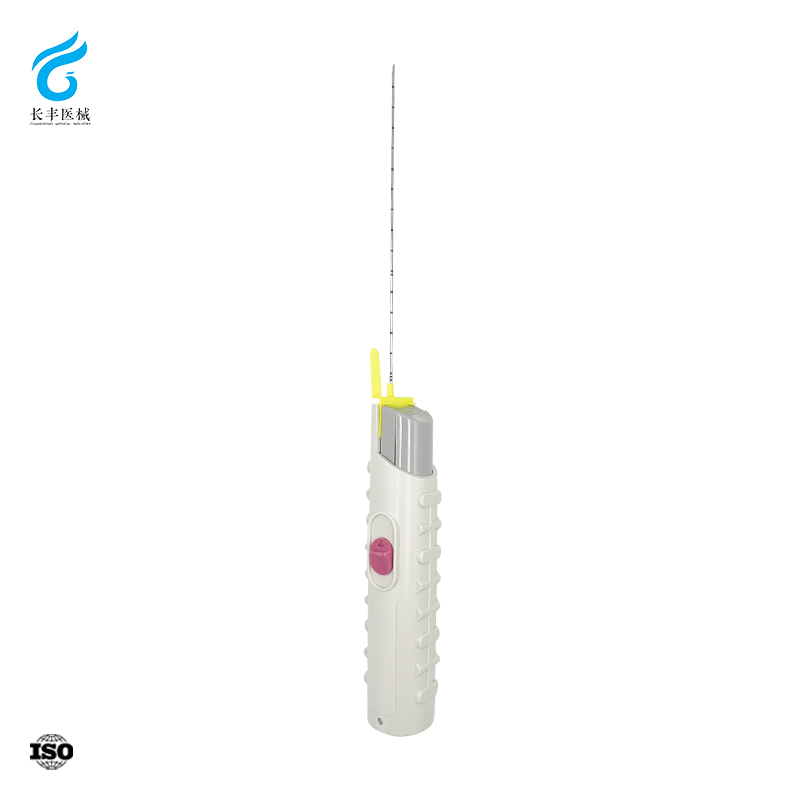
Anatomy-Informed Design: How the Quincke Needle’s Beveled Tip Enables Controlled Dural Penetration
Bevel Geometry and Tissue Interaction: Why Sharpness and Orientation Matter at L3–L4/L4–L5
The Quincke needle’s beveled, cutting tip enables controlled dural penetration by parting—rather than tearing—elastic fibers in the ligamentum flavum and dura mater. This reduces tissue distortion and insertion force. Crucially, bevel orientation determines whether dural fibers are severed or gently separated: when aligned parallel to the longitudinal axis of the dura (typically with the bevel facing laterally or cephalad), the needle spreads fibers apart, preserving structural integrity and lowering the risk of persistent cerebrospinal fluid (CSF) leak. A distinct tactile “pop” signals intradural entry—usually at 5–7 cm depth in adults—providing reliable real-time feedback. Advancing the needle at a 20–40° cranial inclination aligns it with the interlaminar window, avoiding bony contact and supporting smooth, safe passage through layered anatomy.
Biomechanical Advantage: Efficient CSF Access vs. Risk of Dural Tear or Root Trauma
The Quincke needle’s cutting geometry delivers rapid, clean dural puncture with immediate CSF return—streamlining diagnosis and reducing procedural time. Lower insertion force minimizes patient discomfort and needle deflection. Yet this efficiency carries inherent trade-offs: the sharp bevel creates a linear dural slit that may not seal spontaneously, increasing the risk of post-lumbar puncture headache (PLPH) and persistent leak. If the bevel contacts a nerve root—especially with steep angulation or anatomical variation—it may cause transient radicular pain or direct trauma. These risks are mitigated through disciplined technique: careful depth control, real-time assessment of stylet resistance and fluid column behavior, and reliance on the needle’s tactile feedback. In skilled hands, the Quincke’s sharpness is not a hazard but a precision instrument for efficient, controlled access.
Clinical Technique: Optimizing Quincke Needle Use for Reliable Lumbar Puncture Success
Step-by-Step Insertion Protocol: Landmarking, Angle, Depth, and Real-Time CSF Feedback
Begin with optimal positioning—lateral decubitus or seated with full lumbar flexion—to maximize interspinous space width. Palpate the iliac crests to identify the L3–L4 or L4–L5 interspace. After skin antisepsis and local anesthetic infiltration, insert the needle midline with the bevel oriented parallel to dural fibers (commonly cephalad or lateral). Advance at a 10–15° cephalad angle, directing toward the umbilicus. Evidence supports the aspiration technique: applying continuous negative pressure during advancement significantly improves first-attempt success. A 2020 prospective study found that using a 27G Quincke-type needle with aspiration achieved a 93.3% first-attempt dural puncture rate—versus 72.1% with the conventional “popping” method (P=0.019)—and reduced needle withdrawals (8.9% vs. 30.2%) and total procedure time. Aspiration also yielded shallower puncture depths, suggesting earlier detection of subarachnoid entry. Once CSF appears, flow should be free and spontaneous; if sluggish, rotating the needle 90° may optimize bevel alignment with dural fiber orientation.
Troubleshooting Common Failures: Dry Tap, Bloody Tap, and Positional Adjustments with Quincke Needle
A dry tap often reflects inadvertent nerve root contact or extradural placement against the dura. Slight withdrawal and re-advancement with minor angular adjustment frequently resolves this—particularly when guided by aspiration feedback. The same 2020 study confirmed that the popping group required more attempts and deeper penetrations, reinforcing aspiration’s role in avoiding false-depth misinterpretation. A bloody tap usually indicates venous plexus puncture; if CSF does not clear within 3–5 mL, withdraw and reinsert at an alternate interspace. In patients with obesity, degenerative spine disease, or limited mobility, increasing hip flexion or adding gentle lateral tilt can improve interspinous access. The Quincke needle’s sharp bevel—combined with aspiration—enhances discrimination between true subarachnoid entry and extradural placement, reducing unnecessary passes and associated complications.
Evidence-Based Comparison: Quincke vs. Atraumatic Needles in Diagnostic Lumbar Puncture
Post-Lumbar Puncture Headache (PLPH) Rates: Quantifying the Trade-Off Between Ease of Use and Complication Risk
Quincke needles consistently correlate with higher PLPH rates than atraumatic (pencil-point) needles—a difference rooted in dural wound morphology. A 2017 Cochrane review concluded that non-cutting tips reduce PLPH by approximately 60–70% across populations. The Quincke bevel slices dural fibers, creating a larger, irregular defect that delays sealing and promotes CSF leakage. In contrast, pencil-point tips separate fibers without cutting, enabling faster closure.
| Needle Type | Typical PLPH Rate (22G) | Mechanism of Dural Puncture |
|---|---|---|
| Quincke (cutting) | 24–36% | Slices through fibers, leaves gaping defect |
| Atraumatic (pencil-point) | 2–12% | Separates fibers without cutting, faster closure |
Despite this disparity, the Quincke needle remains clinically valuable: its reliable tactile feedback and immediate CSF flashback reduce the number of attempts—an independent risk factor for PLPH. Thus, a single, well-executed Quincke puncture may carry lower overall risk than multiple failed passes with an atraumatic needle in challenging anatomy.
When Quincke Still Wins: Indications Favoring Traditional Needle Use (e.g., Obesity, Scarred Anatomy, Low CSF Pressure)
The Quincke needle’s advantages become decisive in specific clinical contexts:
- Obesity (BMI > 35): Deep soft-tissue layers dampen tactile feedback for atraumatic needles, while the Quincke’s sharp bevel delivers a consistent “pop” and robust CSF return.
- Scarred or calcified anatomy: Fibrosis, ossification, or prior spinal surgery impedes blunt-tip advancement; the Quincke cuts through adhesions with less force, reducing the risk of cauda equina injury from excessive pressure.
- Low CSF-pressure states: In spontaneous intracranial hypotension or severe dehydration, minimal CSF volume compromises atraumatic needle detection—yet the Quincke reliably confirms intrathecal placement via immediate dural penetration.
- Urgent or high-stakes scenarios: In suspected bacterial meningitis or encephalitis, especially with agitation or altered mental status, the Quincke’s one-step success lowers diagnostic delay and procedural failure.
These indications reflect a pragmatic, evidence-informed balance: when technical difficulty threatens safety or timeliness, the Quincke needle’s predictable performance justifies its higher baseline PLPH risk.
Practical Selection Guide: Matching Quincke Needle Specifications to Patient and Procedural Needs
Selecting the right Quincke needle requires aligning specifications with patient anatomy and procedural intent. The following table consolidates key decision points for bedside use.
| Specification | Common Options | Best Suited For | Key Consideration |
|---|---|---|---|
| Gauge | 22 G, 25 G, 27 G | 22 G for therapeutic drainage or low‑pressure states; 25–27 G for diagnostic LP | Higher gauge (smaller diameter) reduces PLPH risk but slows CSF flow and compromises manometry accuracy |
| Length | 90 mm (3.5 in), 120 mm, 150 mm | 90 mm for average‑weight adults; 120–150 mm for obese or heavily muscled patients | Too short increases dry tap risk; too long raises bending and nerve contact risk |
| Bevel orientation | Parallel to dural fibres | All patients | Cephalad or lateral orientation spreads fibers, minimizing CSF leak and improving seal integrity |
| Cutting‑tip design | Sharp, beveled Quincke edge | Fibrotic or ossified interspinous ligaments, prior spinal hardware, scarred anatomy | The cutting tip advances more reliably through dense tissue than pencil‑point alternatives, saving time |
When needle selection accounts for habitus, pathology, and procedural goals—whether for opening pressure measurement, cytology, or therapeutic CSF removal—the likelihood of success rises, and complication risk falls. Thoughtful Quincke needle use transforms lumbar puncture from a routine skill into a targeted, anatomy-informed intervention.
FAQs
Why does bevel orientation matter during lumbar punctures?
Bevel orientation determines whether dural fibers are severed or separated. Proper alignment parallel to dural fibers can reduce tissue damage and minimize the risk of cerebrospinal fluid (CSF) leakage.
How does the Quincke needle compare to atraumatic needles in terms of post-lumbar puncture headache risk?
The Quincke needle is associated with higher rates of post-lumbar puncture headache (PLPH), as its cutting bevel creates a larger dural defect compared to atraumatic needles that separate fibers without slicing.
What gauge is most commonly used for diagnostic lumbar punctures?
For diagnostic lumbar punctures, gauges such as 25G or 27G are preferred as they reduce PLPH risk. However, smaller diameters may slow cerebrospinal fluid collection.
How does obesity affect lumbar puncture technique with Quincke needles?
In obese patients, deeper soft-tissue layers can dampen tactile feedback. Quincke needles are advantageous in such cases due to their sharp bevel that reliably signals dural penetration.
What should be done if a dry or bloody tap occurs?
A dry tap may require slight withdrawal and re-advancement of the needle with angular adjustments. A bloody tap often necessitates re-insertion at an alternate interspace if cerebrospinal fluid does not clear within 3–5 mL.
Table of Contents
- Anatomy-Informed Design: How the Quincke Needle’s Beveled Tip Enables Controlled Dural Penetration
- Clinical Technique: Optimizing Quincke Needle Use for Reliable Lumbar Puncture Success
- Evidence-Based Comparison: Quincke vs. Atraumatic Needles in Diagnostic Lumbar Puncture
- Practical Selection Guide: Matching Quincke Needle Specifications to Patient and Procedural Needs
-
FAQs
- Why does bevel orientation matter during lumbar punctures?
- How does the Quincke needle compare to atraumatic needles in terms of post-lumbar puncture headache risk?
- What gauge is most commonly used for diagnostic lumbar punctures?
- How does obesity affect lumbar puncture technique with Quincke needles?
- What should be done if a dry or bloody tap occurs?