
 EN
EN
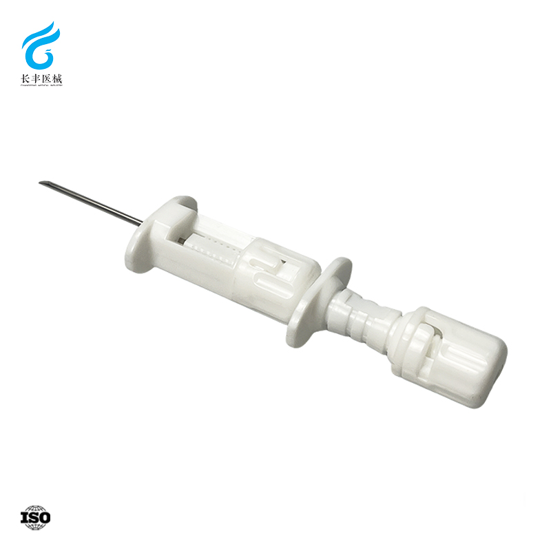
Clinical Indications for Breast Puncture Needle Use
BI-RADS 4/5 Lesions as Primary Triggers for Breast Puncture Needle Intervention
BI-RADS (Breast Imaging Reporting and Data System) category 4 and 5 lesions are suspicious findings on mammography or ultrasound that mandate tissue diagnosis. A breast puncture needle biopsy is the minimally invasive frontline method to obtain a histologic sample. As noted by the American College of Radiology, BI-RADS 4 lesions carry a malignancy risk ranging from 2% to over 95% depending on subcategory, while BI-RADS 5 lesions have a positive predictive value exceeding 95%. This wide probability range underscores the need for precise, image-guided sampling. Core needle biopsy achieves diagnostic accuracy comparable to surgical excision and significantly reduces the need for open procedures. Performed under image guidance—typically in an outpatient setting—it carries a low complication rate. By enabling rapid histologic analysis, this approach helps clinicians confirm or rule out malignancy without delay, supporting timely treatment decisions and reducing patient anxiety. Thus, BI-RADS 4/5 findings remain the primary clinical trigger for needle-based intervention.
Differentiating Cystic vs. Solid Masses Using Breast Puncture Needle Sampling
Ultrasound features such as anechoic appearance and well-defined borders often suggest a simple cyst—but only needle aspiration can definitively confirm its nature. When a mass is identified on imaging, breast puncture needle sampling provides immediate differentiation between cystic and solid lesions. For suspected simple cysts, a fine needle attached to a syringe is advanced under ultrasound guidance; aspiration of clear fluid with complete collapse of the mass confirms benignity. In such cases, fluid is typically discarded unless it appears bloody or the cyst recurs. Aspiration also offers therapeutic relief for large, painful cysts. If the mass fails to collapse fully—or refills rapidly—a core biopsy is indicated to assess for a solid component. Conversely, solid masses or complex cysts with internal echoes or septations require core needle biopsy to obtain tissue for histopathologic evaluation. This dual capability makes breast puncture needle sampling both diagnostic and therapeutic, avoiding unnecessary surgery for benign cysts while ensuring definitive diagnosis of suspicious solid lesions—all within a single, well-tolerated encounter.
Comparing Breast Puncture Needle Techniques: Accuracy, Yield, and Limitations
The choice of breast puncture needle technique directly influences diagnostic accuracy, tissue adequacy, and the risk of false-negative results. Understanding the strengths and limitations of each method enables clinicians to tailor the procedure to the lesion’s characteristics and the clinical question at hand.
Fine-Needle Aspiration (FNA) of Breast Lesions: Role, Sensitivity, and ROSE-Enhanced Utility
Fine-needle aspiration (FNA) uses a thin-gauge needle to extract cells from palpable or image-detected lesions. It is minimally invasive, cost-effective, and particularly useful for rapidly distinguishing cystic from solid masses. However, FNA’s sensitivity varies widely—typically between 60% and 90%—largely due to operator dependency and sampling error. Rapid on-site evaluation (ROSE), where a cytopathologist assesses the aspirate immediately, markedly improves diagnostic yield: multicentre audit data from 2023 show the insufficient sample rate drops from ~20% to under 5% when ROSE is used. While FNA remains valuable for confirming benign cysts or evaluating recurrent lesions, its inability to preserve tissue architecture limits its utility in the primary diagnosis of suspicious solid masses. Today, FNA is most appropriately reserved for scenarios requiring rapid, low-cost confirmation—or when integrated with ROSE in centres with robust cytopathology support.
Core Needle Biopsy vs. Vacuum-Assisted Breast Puncture Needle: Tissue Adequacy and False-Negative Reduction
Core needle biopsy (CNB) and vacuum-assisted biopsy (VAB) both use larger-gauge needles to obtain tissue cores, preserving histologic architecture and enabling immunohistochemical analysis. The table below summarises key differences in tissue adequacy and diagnostic performance.
| Feature | Core Needle Biopsy (14‑18G) | Vacuum-Assisted Breast Puncture Needle (8‑11G) |
|---|---|---|
| Tissue volume per pass | 1–2 cores, ~20 mm³ | 5–20 cores, considerably larger specimen |
| Lesion targeting | Spring-loaded; may miss small or calcified lesions | Directional, multiple contiguous samples; improves retrieval of microcalcifications |
| False-negative rate | 2–4% (2023 systematic review) | <1% in well-selected calcifications |
| Procedure time | Faster; fewer samples | Slightly longer; requires vacuum console |
| Cost | Lower | Higher equipment cost |
CNB provides adequate tissue for definitive diagnosis in most solid masses, though operator experience and lesion size can influence false-negative rates. VAB, using a vacuum-assisted breast puncture needle, excels in sampling microcalcifications and lesions with indistinct margins—contexts where standard CNB may underestimate disease extent. A 2022 meta-analysis of over 8,000 biopsies found VAB reduced the upgrade rate of atypical ductal hyperplasia to malignancy by 40% compared to CNB. Consequently, VAB is the preferred method for stereotactic biopsy of calcifications and for resolving discordant imaging–pathology findings. Selecting between these techniques requires balancing tissue adequacy, procedural efficiency, cost, and the specific diagnostic challenge.
Image-Guided Breast Puncture Needle Procedures: Best Practices and Technical Considerations
Ultrasound-Guided Breast Puncture Needle Biopsy: Workflow, Real-Time Targeting, and Operator Factors
Ultrasound guidance transforms the breast puncture needle procedure by providing real-time visualization of the needle tip—critical for accurately targeting non-palpable lesions. This visual feedback allows continuous adjustment of needle trajectory, ensuring sampling occurs precisely within the area of suspicion rather than adjacent normal tissue. The standard workflow includes a systematic sweep with a high-frequency linear probe to localize the lesion, followed by an in-plane needle approach that keeps the entire shaft visible throughout advancement. A key advantage is immediate confirmation of needle position inside the target, which directly enhances diagnostic accuracy by minimizing sampling error. However, this precision is operator-dependent and requires dedicated training in hand-eye coordination and probe–needle alignment. A controlled study demonstrated that ultrasound guidance achieved a 96% accuracy rate in avoiding sensitive structures—compared to just 40% with palpation-based methods. While the technique demands more time than blind approaches due to meticulous real-time adjustments, clinical best practice firmly establishes ultrasound-guided breast puncture needle biopsy as the standard of care for sonographically visible lesions, prioritising safety, diagnostic yield, and procedural reliability.
FAQ Section
What are BI-RADS 4 and 5 lesions, and why are they important?
BI-RADS 4 and 5 lesions are mammographic or ultrasound findings with a higher risk of malignancy. BI-RADS 4 lesions have a malignancy risk ranging from 2% to over 95%, while BI-RADS 5 lesions have a positive predictive value of over 95%. These lesions typically warrant a tissue diagnosis, often through breast puncture needle biopsy.
How do needle biopsy techniques confirm benign cysts?
Using a fine needle under ultrasound guidance allows aspirating fluid from suspected cystic lesions. Clear fluid and complete collapse of the cyst confirm benignity. If the cyst refills or contains unusual fluid, further evaluation is required.
What is the difference between core needle biopsy and vacuum-assisted biopsy?
Core needle biopsy uses a spring-loaded device to extract tissue cores, while vacuum-assisted biopsy utilizes a vacuum console for larger tissue retrieval. Vacuum-assisted biopsy is especially suited for microcalcifications and lesions that may not be adequately sampled using core needle biopsy.
Why is ultrasound guidance preferred for targeted biopsies?
Ultrasound guidance provides real-time visualization of the needle tip, enabling precise targeting of suspicious lesions and minimizing errors. This technique is operator-dependent and ensures procedural reliability and safety.